Saline Infusion Sonography: A Comprehensive Guide
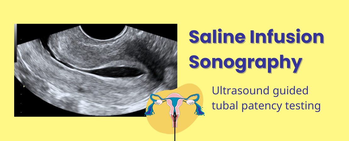
Sonohysterography also called Saline Infusion Sonography (SIS) is a specialized ultrasound technique used to examine the inside of the uterus. By filling the uterus with saline (a saltwater solution), SIS creates clearer ultrasound images, helping doctors diagnose issues like polyps, fibroids, and other uterine abnormalities. This minimally invasive procedure is widely used in gynecology, offering a safer and often more accurate alternative to more invasive tests. Lets find out how SIS aids in detecting abnormalities in the uterus and fallopian tubes, and why it is a valuable tool for diagnosing certain gynecological issues.
What is Saline Infusion Sonography?
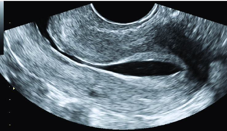
SIS is an ultrasound procedure that uses sterile saline to improve the visibility of the uterine lining. The saline expands the uterus, making it easier to spot abnormalities that might not show up on regular ultrasounds. This method is particularly useful for detecting problems like uterine polyps, fibroids, and scar tissue.
How Does Saline Infusion Sonography Work?
During SIS, a thin tube is inserted into the uterus through the cervix. Saline is then gently infused into the uterus, expanding it and allowing for a clear view of the uterine lining with a transvaginal ultrasound. The saline creates a fluid-filled space that helps distinguish the endometrium from any abnormalities. The procedure is quick, usually taking 15-30 minutes, and is done on an outpatient basis.
Indications for Saline Infusion Sonography
SIS is used to diagnose and evaluate a variety of uterine conditions. Common reasons for performing SIS include:
- Abnormal Uterine Bleeding: Helps identify the cause of unexplained bleeding.
- Infertility Assessments: Checks for uterine abnormalities that could affect fertility.
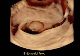
- Endometrial Polyps: Detects benign growths inside the uterus.
- Uterine Fibroids: Identifies non-cancerous tumors in the uterus.
- Intrauterine Adhesions: Evaluates scar tissue within the uterus, often due to previous surgeries or infections.
Preparing for the SIS Procedure
Before undergoing SIS, patients should follow a few simple guidelines:
- Avoid eating heavy meals before the procedure.
- Take any prescribed pain relievers if recommended by your doctor.
- Inform your doctor of any allergies, especially to latex or iodine.
- SIS is usually scheduled just after menstruation ends, when the endometrium is thin and easy to evaluate.
- Women who might be pregnant should not undergo SIS.
The SIS Procedure: Step-by-Step
The SIS procedure is straightforward and typically includes these steps:
- Insertion of the Catheter: A thin, flexible tube is gently inserted into the uterus through the cervix.
- Saline Infusion: Sterile saline is slowly introduced into the uterus through the catheter, expanding the uterine cavity.
- Ultrasound Imaging: A transvaginal ultrasound probe is used to capture images of the uterus, with the saline highlighting any abnormalities.
- Completion: After imaging, the saline is naturally expelled, and the catheter is removed. The entire procedure usually takes less than 30 minutes.
What to Expect During and After the Procedure
During SIS, you might feel some mild cramping or discomfort when the catheter is inserted and as the saline fills the uterus. However, the procedure is generally well-tolerated, and the discomfort is usually brief.
After the procedure, some women experience light spotting and mild cramping, but these symptoms typically resolve within a day. Most patients can resume normal activities immediately. Your doctor may advise you to avoid strenuous exercise or sexual activity for 24 hours to allow your body to recover fully.
Benefits of Saline Infusion Sonography
SIS offers several advantages over other diagnostic methods:
- High Accuracy: SIS provides detailed images that help accurately diagnose uterine abnormalities.
- Minimally Invasive: The procedure is less invasive than surgical alternatives like hysteroscopy.
- Less Painfull: The procedure is less painfully than HSG, an x-ray alternative.
- Cost-Effective: SIS is generally more affordable compared to other imaging techniques, like MRI.
- Quick Recovery: Since SIS is minimally invasive, patients can return to their daily activities almost immediately.
Risks and Complications Associated with SIS
While SIS is generally safe, it does carry some risks:
- Mild Cramping: Some women experience cramping during or after the procedure.
- Spotting: Light bleeding may occur for a day or two after SIS.
- Infection: There is a small risk of infection, which can be minimized by following pre-procedure guidelines.
- Allergic Reaction: Rarely, an allergic reaction to the saline or latex used in the procedure may occur.
If you experience severe pain, heavy bleeding, or symptoms of infection (like fever or foul-smelling discharge) after the procedure, contact your doctor immediately.
SIS vs. Hysteroscopy: A Comparative Analysis
Saline Infusion Sonography (SIS) and hysteroscopy are both used to evaluate the uterine cavity, but they differ in several key ways:
- Invasiveness: SIS is minimally invasive, involving only the insertion of a small catheter and the use of ultrasound. Hysteroscopy, on the other hand, involves inserting a camera through the cervix into the uterus, making it more invasive.
- Diagnostic vs. Operative: SIS is primarily diagnostic, providing detailed images of the uterus. Hysteroscopy can be both diagnostic and operative, allowing for the removal of polyps, fibroids, and other abnormalities during the same procedure.
- Cost and Recovery: SIS is generally less expensive and involves a quicker recovery time compared to hysteroscopy.
- When to Choose: SIS is preferred for initial evaluations and when a non-invasive approach is desired. Hysteroscopy is chosen when there’s a need for both diagnosis and immediate treatment.
Advances in Saline Infusion Sonography
Recent advancements in SIS technology have improved its diagnostic capabilities:


- 3D Imaging: The introduction of 3D ultrasound in SIS allows for even more detailed and accurate visualization of the uterine cavity, helping in better diagnosis and treatment planning.
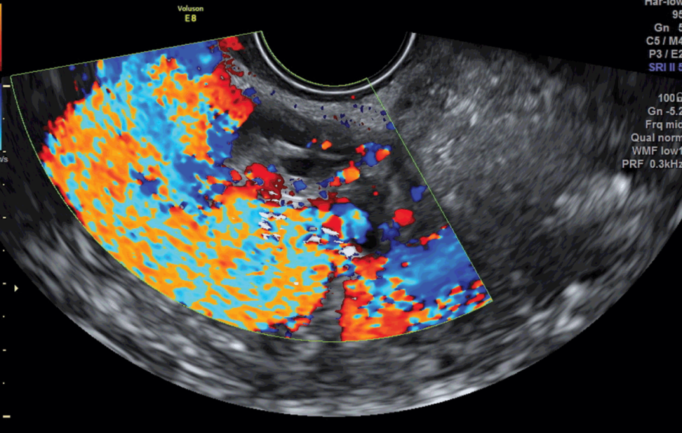
- Enhanced Ultrasound Techniques: Doppler ultrasound, when combined with SIS, can assess blood flow within the uterus, providing additional information about uterine and endometrial health.
- Automated Systems: Newer SIS systems are more automated, reducing the time required for the procedure and improving patient comfort.
These advances make SIS a more powerful tool in diagnosing uterine conditions and tailoring individual treatment plans.
Case Studies and Clinical Applications
SIS has been successfully used in a variety of clinical scenarios:
- Case Study 1: A 35-year-old woman with abnormal uterine bleeding underwent SIS, which revealed a small polyp that was missed on standard ultrasound. Removal of the polyp resolved her symptoms.
- Case Study 2: In a fertility clinic, a 30-year-old woman with unexplained infertility had an SIS that identified intrauterine adhesions. After treatment, she successfully conceived.
- Case Study 3: A postmenopausal woman was referred for SIS after experiencing unexpected bleeding. SIS revealed thickened endometrium, prompting further investigation that led to early detection of endometrial cancer.
These examples highlight the effectiveness of SIS in diagnosing and guiding treatment for various uterine conditions.
Saline Infusion Sonography in Fertility Treatments
SIS plays a crucial role in fertility assessments. It is often used to evaluate the uterine cavity for abnormalities that could affect a woman’s ability to conceive or maintain a pregnancy.
- Uterine Abnormalities: SIS can detect polyps, fibroids, and scar tissue that might interfere with embryo implantation or cause recurrent miscarriages.
- Pre-IVF Assessments: Before in vitro fertilization (IVF), SIS helps ensure that the uterine environment is optimal for embryo transfer.
- Post-Treatment Monitoring: After fertility treatments like hysteroscopic surgery, SIS can be used to confirm that the uterine cavity is clear and ready for pregnancy.
In fertility clinics, SIS is a valuable tool for diagnosing and addressing uterine factors that could impact treatment outcomes.
Patient Experiences and Testimonials
Many patients find SIS to be a straightforward and informative procedure:
- Comfortable Experience: Most patients report that the procedure is quick and causes only mild discomfort.
- Clear Results: Patients appreciate the clear and immediate feedback provided by the ultrasound images, which often help to explain symptoms and guide treatment.
- Reassurance: Many patients feel reassured after SIS, knowing that their uterine cavity has been thoroughly evaluated.
Clinicians often share that patients who undergo SIS are more engaged in their care and better understand their treatment options.
Conclusion
Saline Infusion Sonography is a valuable diagnostic tool in gynecology, offering a clear, non-invasive method for evaluating the uterine cavity. Whether used to investigate abnormal bleeding, assess fertility issues, or monitor treatment outcomes, SIS provides high-quality images that guide effective treatment plans. With advances in imaging technology and its growing role in fertility treatments, SIS continues to be a critical procedure in modern gynecological practice.
As with any medical procedure, it’s essential for patients to discuss the potential benefits and risks with their healthcare provider to ensure that SIS is the right choice for their individual needs.
FAQs About Saline Infusion Sonography
1. Is Saline Infusion Sonography painful?
SIS is generally well-tolerated and causes minimal discomfort. Some women may experience mild cramping during the procedure, but it is typically brief and manageable.
2. How should I prepare for a Saline Infusion Sonography?
Your doctor may advise you to avoid heavy meals before the procedure and to take an over-the-counter pain reliever if needed. It’s important to inform your doctor about any allergies or if there’s a chance you could be pregnant.
3. How long does the SIS procedure take?
The SIS procedure usually takes about 15 to 30 minutes from start to finish. It is performed on an outpatient basis, so you can go home the same day.
4. Are there any risks or side effects associated with SIS?
While SIS is a safe procedure, some risks include mild cramping, light spotting, or, in rare cases, infection. These side effects are usually temporary and resolve quickly.
5. Can SIS be used during fertility treatments?
Yes, SIS is often used in fertility treatments to evaluate the uterine cavity for abnormalities that could affect conception or pregnancy. It helps in diagnosing issues like polyps, fibroids, and adhesions that might impact fertility.
Additional Reading:
PUBMED: Saline infusion sonohysterography
ACOG: Sonohysterography


